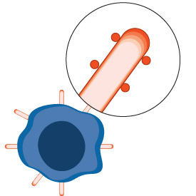
Chimeric antigen receptor (CAR) T therapy is a revolutionary type of adoptive cell immunotherapy, which employs genetic engineering to redirect the specificity of cytotoxic T lymphocytes to a tumor antigen(s) expressed on the surface of tumor cells. CAR-T therapy has shown remarkable success in the treatment of hematologic malignancies and could potentially be leveraged to safely treat solid tumors and diseases of senescence, such as fibrotic liver disease and diabetes.1-3 CAR-T immunotherapies have been described as living therapies that may be efficacious decades after a single infusion.
The CAR-T development workflow is a multi-step process that ensures the development of safe, effective treatments. Cell Signaling Technology® (CST) offers product solutions to aid in preclinical research for target identification and selection, CAR-T cell characterization, and interrogation of patient response to therapy.
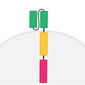
To aid in characterizing CAR-T cells, CST offers innovative and award-winning CAR Linker antibodies that target the G4S and Whitlow linker sequences of scFv-based CARs. Additionally, you can find fluorochrome-conjugated antibodies for CAR-T cell phenotyping and multiple IHC-validated monoclonal antibodies to analyze CAR targets in tissues. With a growing portfolio of related products, CST supports your CAR-T research, every step of the way.
 |
 |
 |
 |
|
Step 1: |
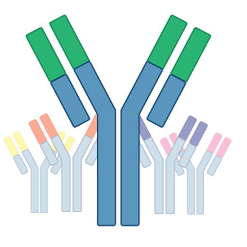
Step 2: scFv Screening and Optimization |
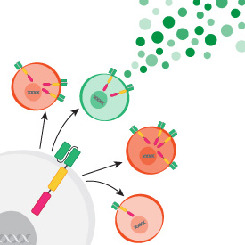
Step 3: CAR Transgene Construction |
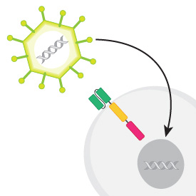
Step 4: CAR Transgene Delivery |
 |
 |
 |
|
|
Step 5: |
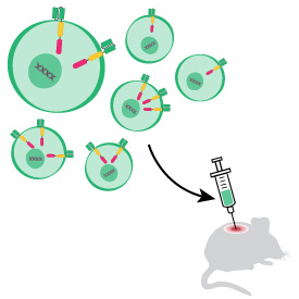
Step 6: Preclinical In Vivo Assays |
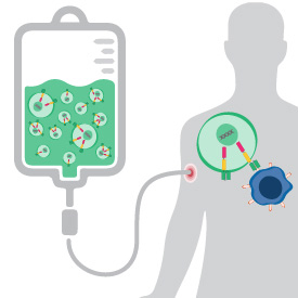
Step 7: Patient Selection and Monitoring |
The ultimate goal of tumor antigen discovery and validation is to find tumor surface antigens that are expressed on the majority of tumor cells; display an expression pattern that minimizes risk of on-target, off-tumor toxicity; and are expressed stably so target loss will not render the treatment ineffective. Identifying a new tumor antigen is challenging enough without having to worry about whether the antibodies you’re using are specific or are of sufficient sensitivity to detect potential tumor antigens. You need to use reliable antibodies that you can trust to detect the tumor target protein accurately every time. CST antibodies undergo rigorous in-house application-specific validation, giving you confidence that your detection reagents are specific, reliable, and high-affinity.
Start with highly specific antibodies to detect known tumor antigens in your sample: |
|
|---|---|
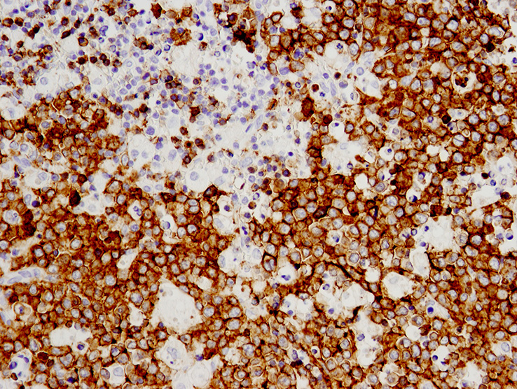
Immunohistochemical analysis of paraffin-embedded human lymphoma using CD19 (Intracellular Domain) (D4V4B) XP® Rabbit mAb #90176. 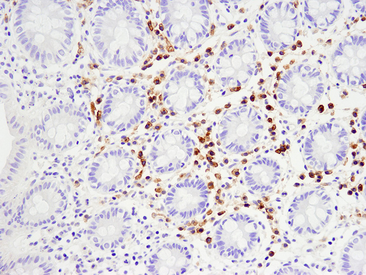
Immunohistochemical analysis of paraffin-embedded human normal colon using TNFRSF17/BCMA (E6D7B) Rabbit mAb #88183. |
|
IHC-validated antibodies or flow cytometry-validated antibody conjugates: |
|

Flow cytometric analysis of HeLa cells (blue) and KARPAS-299 cells (green) using TNFRSF8/CD30 (E7E4D) XP® Rabbit mAb (Alexa Fluor® 488 Conjugate) (solid lines) or concentration-matched Rabbit (DA1E) mAb IgG XP® Isotype Control (Alexa Fluor® 488 Conjugate) #2975 (dashed lines). KARPAS cell line source: Dr. Abraham Karpas at the University of Cambridge. 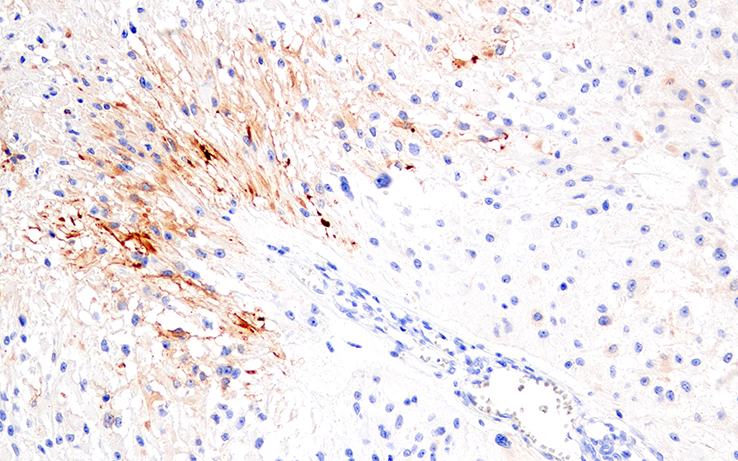
Immunohistochemical analysis of paraffin-embedded human glioblastoma multiforme using IL-13RA2/CD213a2 (E7U7B) Rabbit mAb #85677. |
|
Proliferation AssaysThe ability of a CAR-T cell to proliferate in response to engaging a tumor antigen is an important readout of CAR-T cell function.4 Evaluate your CAR-T cells' ability to propagate with a CST proliferation assay. The Cell Proliferation Tracer Kits use flow cytometry to monitor a fluorescent tracer dye that gets diluted with each cell division. |
|
|---|---|

Cell division tracking in live Jurkat cells over the course of 4 days (d0-d3). Cells were labeled with the Cell Proliferation Tracer Kit (Fluorometric, Violet 450) #48444 on day 0, and analyzed by flow cytometry each day. Each successively dimmer peak represents one cell division. Unstained cells are represented by the unshaded peak. 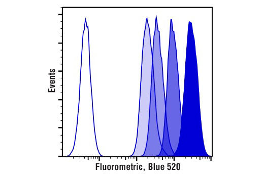
Cell division tracking in live Jurkat cells over the course of 4 days (d0-d3). Cells were labeled with the Cell Proliferation Tracer Kit (Fluorometric, Blue 520) #53452 on day 0, and analyzed by flow cytometry each day. Each successively dimmer peak represents one cell division. Unstained cells are represented by the unshaded peak. |
|
Cytotoxicity AssaysCytolytic assays are routinely used to determine immune effector cell function. Interrogate cell-mediated cytotoxicity with a sensitive kit that does not employ radioisotopes. |
|
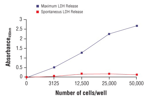
Determination of Optimum Cell Number for LDH Cytotoxicity Assay: HeLa cells were seeded into a 96-well plate at varying densities using media containing 10% FBS. After overnight incubation, the cells were replaced with serum-free media and then treated with either Assay Buffer (Spontaneous LDH Release) or 10% Triton™ X-100 solution (Maximum LDH Release). After treatment, the medium was removed and placed into a new 96-well plate. The amount of LDH released into the medium was determined using the LDH Cytotoxicity Assay Kit protocol. |
|
Explore the role of the immune system in oncology using immunophenotyping antibodies. Applications like immunohistochemistry (IHC) and flow cytometry allow for multiplex monitoring of cell populations and both require stringent antibody validation principles.Phenotype T cell subsets to understand which will be most efficacious for engineering or phenotype the CAR-T cells to understand what effect the CAR has on phenotype and function.Explore a comprehensive catalog of markers for immune phenotype and function optimized for tissue or liquid biopsy sample analysis. |
|
|---|---|
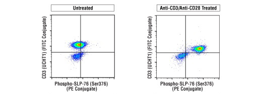
Flow cytometric analysis of human peripheral blood mononuclear cells, untreated (left) or treated with cross-linked anti-CD3 plus anti-CD28 (10 μg/ml each, 15 min; right), using Phospho-SLP-76 (Ser376) (E3G9U) XP® Rabbit mAb (PE Conjugate) #76143 and co-stained with CD3 (UCHT1) Mouse mAb (FITC Conjugate) #86774. 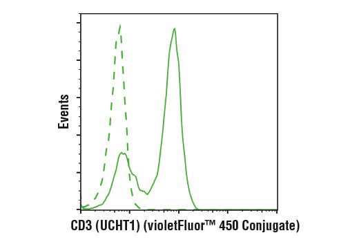
Flow cytometric analysis of live human peripheral blood mononuclear cells using CD3 (UCHT1) Mouse mAb (violetFluor™ 450 Conjugate) #61347 (solid line) compared to concentration-matched Mouse (MOPC-21) mAb IgG1 Isotype Control (violetFluor™ 450 Conjugate) #40282 (dashed line). |
|
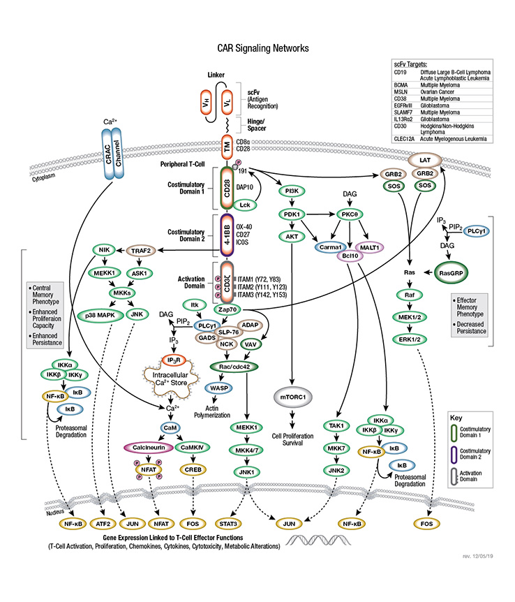
Monitor the activation state of multiple proteins related to T-cell behavior to optimize CAR functionality. CST offers a diverse array of antibodies against targets in relevant signaling pathways, enabling you to evaluate downstream outcomes and better understand CAR-T cell behavior. Check out the CAR Signaling Network pathway and other pathways by research area.To globally interrogate the phospho-proteome that is engaged by CARs, CST offers Proteomics Analytical Services for qualitative and quantitative proteomic profiling. Our experts are ready to partner with you from project planning to the delivery of a comprehensive data package. Explore our Proteomics Analytical Services offerings. |
|
|---|---|
 |
|
Don’t see the flow cytometry antibody conjugate you need? Not a problem! Take advantage of our expertise in conjugating antibodies. We’ll conjugate the antibody you need to your fluorochrome of choice so you have the flexibility to design the assay you want. Custom antibody conjugations are validated, optimized, and tested for stability using the same standards we apply to all CST antibodies.
Order your custom antibody conjugate here!
Carrier-free and customized formulations of your favorite antibody are available so you can perform CAR-T characterization assays free of BSA, glycerol, or azide. Your antibody of choice is offered in PBS for optimal use in applications such as:
Explore our list of ready-to-use, carrier-free antibodies.
Or request a custom formulation of CST catalog antibodies.
| CAR Protein Detection Tools | Features |
|---|---|
|
G4S monoclonal antibodies |
|
|
Whitlow/218 monoclonal antibodies |
|
|
Protein L |
|
|
Epitope tag antibodies (Myc, FLAG, and HA) |
|
|
Surrogate markers of CAR expression (GFP, TagBFP, LNGFR) |
|
|
Anti-idiotype monoclonal antibodies |
|
|
Recombinant CAR target antigen |
|
For Research Use Only. Not For Any Diagnostic of Therapeutic Use.
Cell Signaling Technology is a trademark of Cell Signaling Technology, Inc.
XP is a registered trademark of Cell Signaling Technology, Inc.
All other trademarks are the property of their respective owners. Visit cellsignal.com/trademarks for more information.